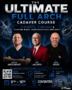
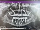
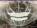
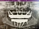

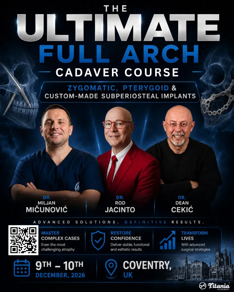
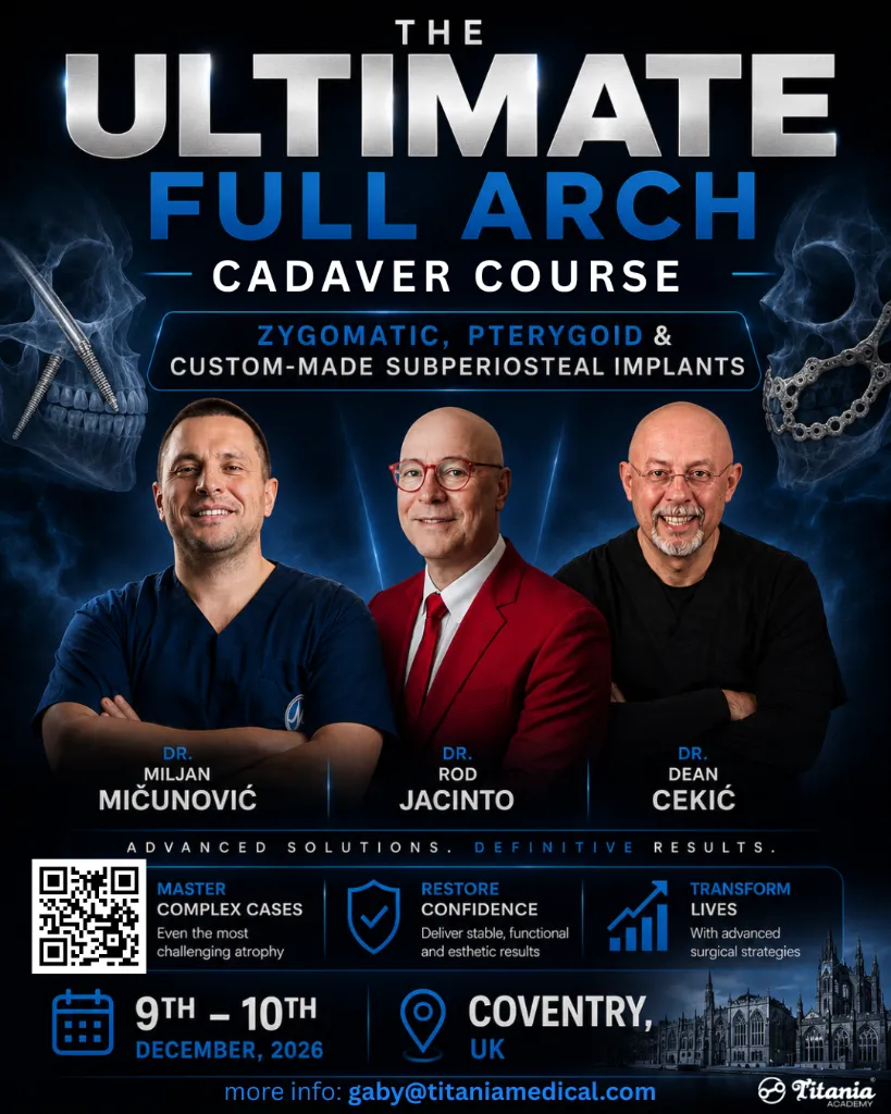

The Ultimate Full Arch Cadaver Course | Zygomatic, Pterygoid & Custom-made Subperiosteal Implants || SPECIAL TUITION UNTIL 27TH SEPTEMBER!!
SPECIAL TUITION RATE
Available for registrations until 27 September
Places are limited, and with Dr Miljan's courses traditionally filling quickly, we strongly recommend securing your place early.
If you are serious about advancing your approach to complex full-arch cases, this is an opportunity you won't want to miss.
Lecturers:
Dr Miljan Mičunović
Dr Dean Cekić
Dr Rod Jacinto (guest speaker)
9th & 10th December, 2026
Coventry – UK
Course Overview
This intensive, two-day educational programme is specifically engineered for advanced clinicians aiming to master full-arch rehabilitation of the severely atrophic maxilla. Standard implant therapies often fall short in cases of extreme bone resorption; this course addresses those limitations by delivering a comprehensive, multi-modal surgical curriculum.
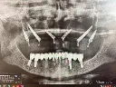
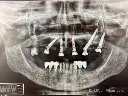
Participants will explore and gain practical proficiency in the three premier, graftless anchorage strategies:
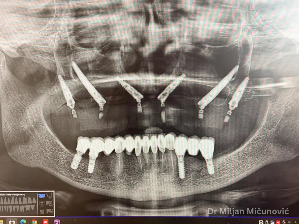
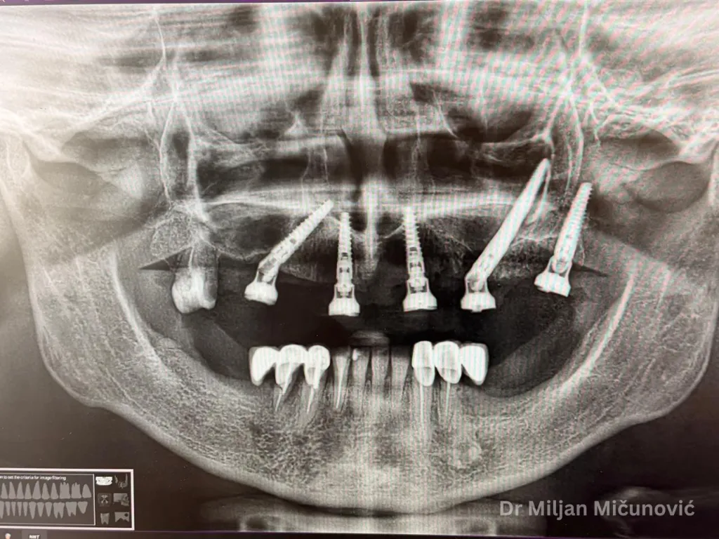
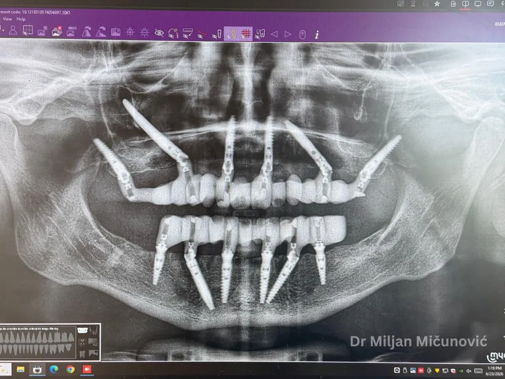
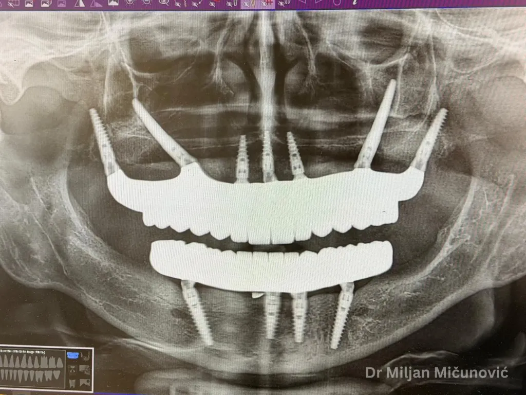
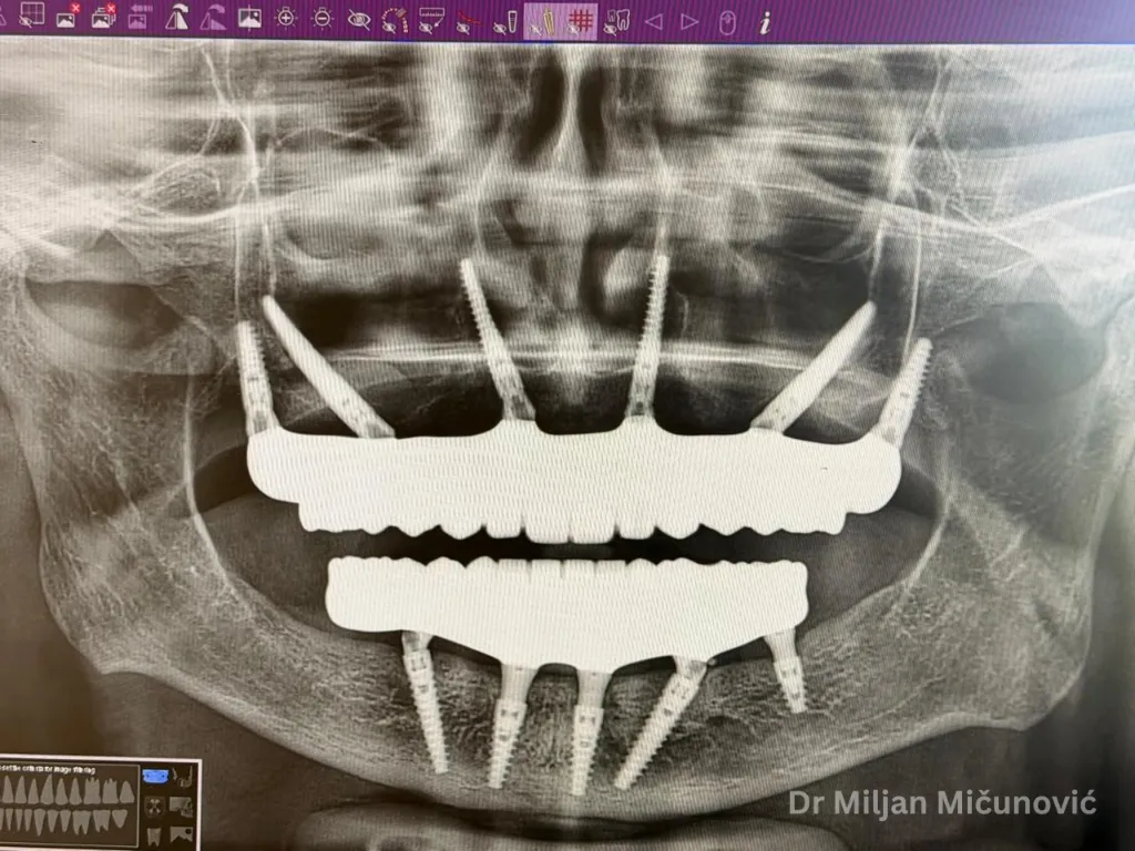
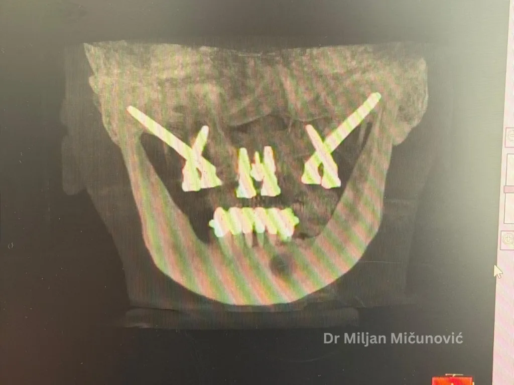
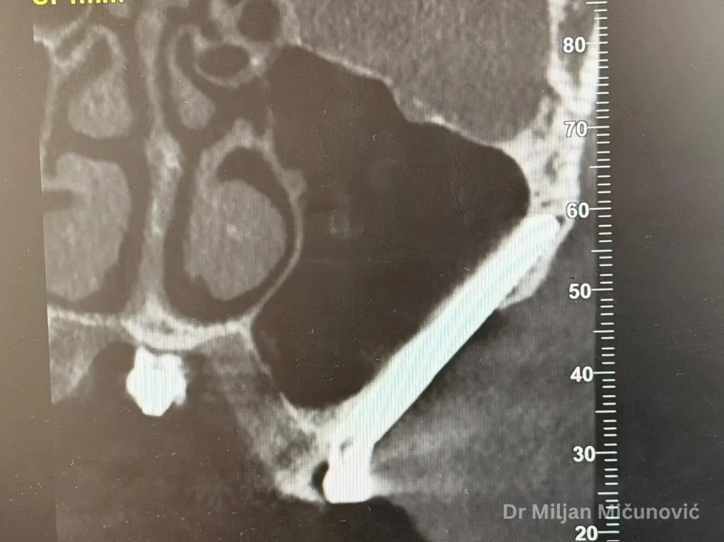
- Zygomatic Implants: Utilising cortical bone of the zygomatic arch for reliable anterior-midfacial anchorage.
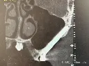
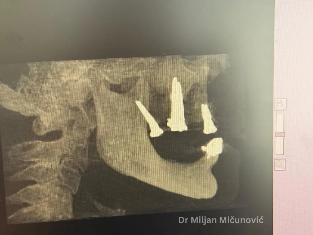
- Pterygoid Implants: Accessing the pterygomaxillary suture to secure posterior cortical anchorage and avoid complex sinus grafting.
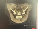
- Patient-Specific Custom Subperiosteal Implants: Leveraging cutting-edge CAD/CAM digital workflows to design and place tailor-made, direct-to-bone titanium frameworks.
Through a blend of evidence-based lecture modules, hands-on synthetic workshops, and personal hands-on cadaver surgical training, delegates will acquire the critical diagnostic, planning, and execution skills required to confidently deliver predictable full-arch rehabilitation outcomes.
Why Attend?
This unique Master Class provides delegates with the opportunity to gain hands-on experience in advanced zygomatic and pterygoid implant surgery, alongside the complete digital workflow for patient-specific subperiosteal implants—from diagnosis and treatment planning through to surgical placement.
Each delegate will be assigned an individual cadaver specimen. A CBCT scan will be acquired for each specimen, enabling the design and manufacture of a patient-specific subperiosteal implant prior to the course.
Delegates will perform advanced surgical procedures focusing on zygomatic and pterygoid implant placement, gaining practical experience with these graftless full-arch rehabilitation techniques before proceeding to the placement of their custom-designed, patient-specific subperiosteal implant.
Unique Learning Experience
Master the three advanced solutions for the Atrophic Maxilla
- Zygomatic Implants: Learn indications, planning, anatomy, surgical protocols, and prosthetic considerations.
- Pterygoid Implants: Understand posterior anchorage techniques, case selection, and surgical execution.
- Patient-Specific Subperiosteal Implants: Master the complete digital workflow from CBCT planning and implant design to surgical placement.
Hands-On Training with Patient-Specific Models
Every participant will work with a 3D-printed anatomical model produced from their own patient's CBCT scan, creating a truly patient-specific learning experience. Delegates will gain hands-on experience in zygomatic and pterygoid implant placement, as well as the surgical placement of a custom-designed, patient-specific subperiosteal implant..
Day 1 – Participants will practise zygomatic and pterygoid implant placement on anatomical models. They will also rehearse the placement of their custom-designed subperiosteal implant on a 3D-printed model replicating their own patient's anatomy, allowing hands-on familiarisation before the live surgery.
Day 2 – Live Surgery: Participants perform the actual surgical procedure, placing the zygomatic and pterygoid implants, followed by the patient-specific subperiosteal implant designed specifically for their patient's case.
This unique two-day format bridges simulation and live application, ensuring each clinician enters surgery having already practiced on an exact replica of their patient's anatomy.